Useful notes for medical students
Click here to view an earlier site with useful notes, unfortunately some images have been lost, however all the text is still present.

Diabetic retinopathy
Background Diabetic Retinopathy:
-
Microaneurysms
-
Dot and Blot hemorrhages
-
Hard exudates (Lipid deposition)
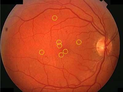
FIGURE 1 MICROANEURYSMS HIGHLIGHTED BY YELLOW RINGS
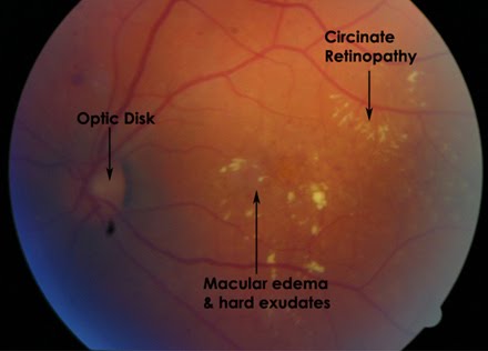
Figure 2 Hard exudates
Pre Proliferative Diabetic Retinopathy
-
IRMA
-
FLAME HEMORRHAGES
-
VENOUS BEADING
-
OMEGA LOOPING OF VENULES
-
COTTON WOOL SPOTS
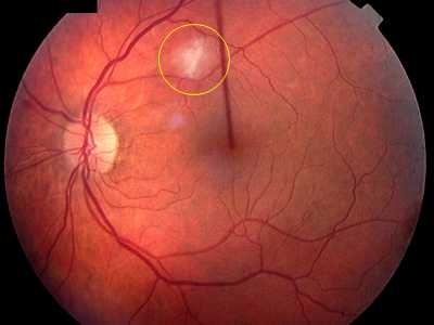
FIGURE 3 COTTON WOOL SPOTS HIGHLIGHTED IN THE YELLOW CIRCLE
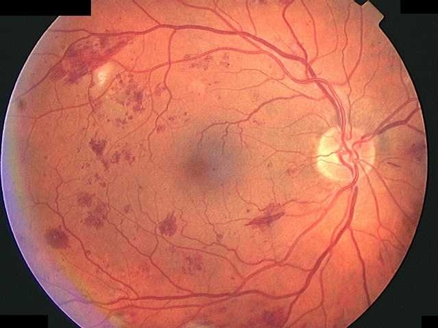
FIGURE 4 Showing IRMA, Cotton wool spots and Flame hemorrhages.
PROLIFERATIVE DIABETIC RETINOPATHY
-
Neovasculariation of disc (NVD)
-
Neovasculariation elsewhere (NVE)
-
Vitreous Hemorrhage
-
Tractional retinal detachment
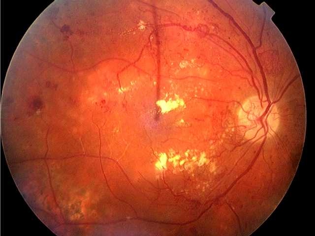
FIGURE 5 Neovasculariation of disc (NVD) and elsewhere (NVE) plus hard exudates within the macular region.
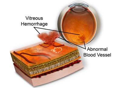
Figure 6 illustrates new abnormal retinal blood vessels which are fragile and may bleed.
References:
-
www.diabeticretinopathy.org.uk
-
Michael Colucciello, MD Diabetic retinopathy Control of systemic factors preserves vision Vol 116 / no 1 / July 2004 / postgraduate medicine
-
Javitt JC, Canner JK, Sommer A. Cost effectiveness of current approaches to the control of retinopathy in type I diabetics. Ophthalmology 1989;96(2):255-64
-
Caro JJ, Ward AJ, O'Brien JA. Lifetime costs of complications resulting from type 2 diabetes in the US. Diabetes Care 2002;25(3):476-81
-
Bresnick GH, Mukamel DB, Dickinson JC, et al. A screening approach to the surveillance of patients with diabetes for the presence of vision-threatening retinopathy. Ophthalmology 2000;107(1):19-24
-
Ferris FL 3rd, Davis MD, Aiello LM. Treatment of diabetic retinopathy. N Engl J Med 1999;341(9):667-78 The effect of intensive treatment of diabetes on the development and progression of long-term complications in insulin-dependent diabetes mellitus.
-
The Diabetes Control and Complications Trial Research Group. N Engl J Med 1993;29(14):977-86
-
Klein R, Klein BE, Moss SE, et al. The Wisconsin Epidemiologic Study of Diabetic Retinopathy: XVII. The 14-year incidence and progression of diabetic retinopathy and associated risk factors in type 1 diabetes. Ophthalmology 1998;105(10):1801-15
-
Aiello LM. Perspectives on diabetic retinopathy. Am J Ophthalmol 2003;136(1):122-35
-
Chaturvedi N, Sjolie AK, Stephenson JM, et al. Effect of lisinopril on progression of retinopathy in normotensive people with type 1 diabetes. The EUCLID Study Group. EURODIAB Controlled Trial of Lisinopril in Insulin-Dependent Diabetes Mellitus. Lancet 1998;351(9095):28-31
-
Chaturvedi N. Modulation of the renin-angiotensin system and retinopathy. Heart 2000;84(Suppl 1):i29-31
-
Chew EY, Klein ML, Ferris FL 3rd, et al. Association of elevated serum lipid levels with retinal hard exudate in diabetic retinopathy. Early Treatment Diabetic Retinopathy Study (ETDRS) Report 22. Arch Ophthalmol 1996;114(9):1079-84